
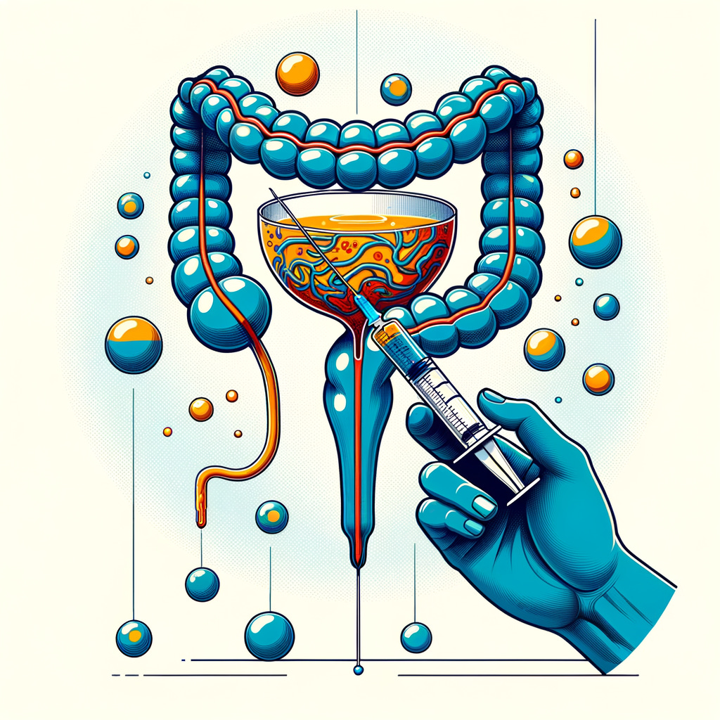
13. Guidelines for Intratumoral Injection of Chlorine Dioxide in Bladder Cancer

13.1 Indications
13.1.1 Bladder cancer patients unsuitable or unwilling to undergo surgery.
13.1.2 Regional lymph node or distant metastasis.
13.1.3 Recurrence post-surgery or post-chemotherapy/radiotherapy.
13.1.4 No active diabetes, tuberculosis, or other conditions.
13.1.5 Normal bleeding and clotting times.
13.2 Contraindications
13.2.1 Cachexia or active diabetes, tuberculosis, with signs of heart, lung, liver, or kidney failure.
13.2.2 Platelet count < 60×10⁹/L.
13.2.3 KPS score ≤ 50.
13.3 Puncture Needle and Auxiliary Instruments
Use a 23G×15.0 cm or 25G×9.0 cm puncture needle, 10–20 ml high-pressure syringe, ultrasound or CT machine.
13.4 Pre-treatment Preparation
13.4.1 Complete necessary examinations:
Routine blood, urine, and stool tests.
Liver and kidney function tests.
Tumor markers: CEA, TPA (bladder cancer tissue polypeptide antigen).
13.4.2 Discuss the procedure with the patient, obtain consent. Instruct on:
Bladder filling by retaining urine before treatment.
Possible complications like fever, local pain, bladder perforation, pelvic inflammation, hematuria.
13.4.3 Administer intramuscularly butorphanol 0.1g, antihemorrhagic agent 1KU, ondansetron 4-8mg, and optionally diazepam 5-10mg, 15 minutes before treatment.
13.5 Treatment Procedure
13.5.1 Under ultrasound or CT guidance, identify the puncture point in the lower abdomen, measure the distance from skin to tumor center, and confirm needle direction.
13.5.2 Disinfect the puncture site skin, wear sterile gloves, and use a sterile drape.
13.5.3 Use 2% lidocaine solution for local anesthesia, stabilize the skin with the left hand and insert the needle with the right hand into the tumor center, then remove the needle core and connect a high-pressure syringe to inject the chlorine dioxide solution. For tumors ≤4 cm, use a single-point injection with a dose of 30% of the tumor volume (ml); for tumors >4 cm, use multi-point injections based on tumor segment volume (cm).
13.5.4 The entire procedure is guided by ultrasound or CT, observing drug distribution within the tumor until saturation, then remove the needle.
13.5.5 Cover the puncture site with sterile gauze and secure with tape.
13.5.6 Observe the patient for 5–10 minutes before transferring back to the ward.
13.6 Post-treatment Care
13.6.1 Within the first week, monitor for fever, local pain, signs of peritonitis or pelvic inflammation, hematuria, and urinary irritation, and provide symptomatic treatment as necessary. If infection is suspected, administer antibiotics and check blood and urine. After one week, reassess blood, urine, liver, and kidney functions, noting symptom relief.
13.6.2 For multiple treatments, allow a 7-day interval. Post-treatment observations remain the same as after the first session. At the end of the treatment cycle, assess peripheral blood T lymphocyte subsets (CD4, CD8) and perform ultrasound or CT to monitor immune status and tumor changes.