

Toy Poodle follow-up, 27 August 2026 — 18 days after the first intratumoral chlorine dioxide ablation
This is a follow-up to the 9 August 2026 case report, “Intratumoral Chlorine Dioxide Ablation of a Recurrent Oral Melanoma in a 12-Year-Old Toy Poodle,” which documented a rapidly growing recurrent oral melanoma of approximately 28 mL and its first ultrasound-guided intratumoral treatment.
Eighteen days later: smaller overall, but not simply resolved
On 27 August, the dog returned for a second injection. Before the first procedure on 9 August, the tumour volume had been estimated at approximately 28 mL. Before today’s second procedure, it was approximately 15 mL. The gross mass was therefore substantially smaller than at baseline, although it had begun to enlarge again after the initial reduction.
The interval appearance was also structurally different. Multiple necrotic cavities were present within the mass. This matters procedurally: the second treatment was not an injection into the same solid geometry seen on Day 1. It was an injection into a smaller, irregular, partially cavitated and relatively loose tumour.
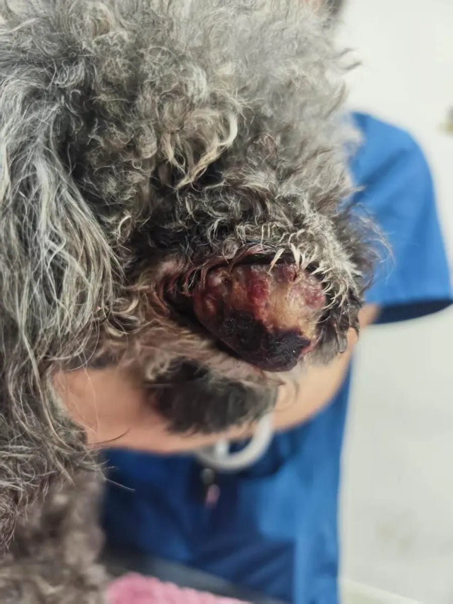
Figure 1. 9 August 2026, before the first injection. Estimated tumour volume: approximately 28 mL.
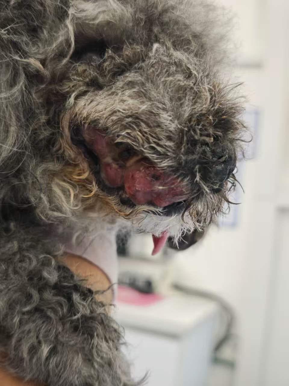
Figure 2. 27 August 2026, before the second injection. Estimated tumour volume: approximately 15 mL; the mass had decreased after the first treatment and subsequently shown renewed growth.
The second procedure
After anaesthesia, the tumour was exposed for direct examination. Areas of cavitation and necrotic material were visible within the mass. A total of approximately 15 mL was injected during the procedure. Because the tumour was loose and contained open or partially open necrotic spaces, substantial leakage occurred. Roughly half of the delivered liquid was estimated to have escaped rather than remaining within viable tumour tissue; the effective retained dose was therefore estimated at approximately 5–7 mL.
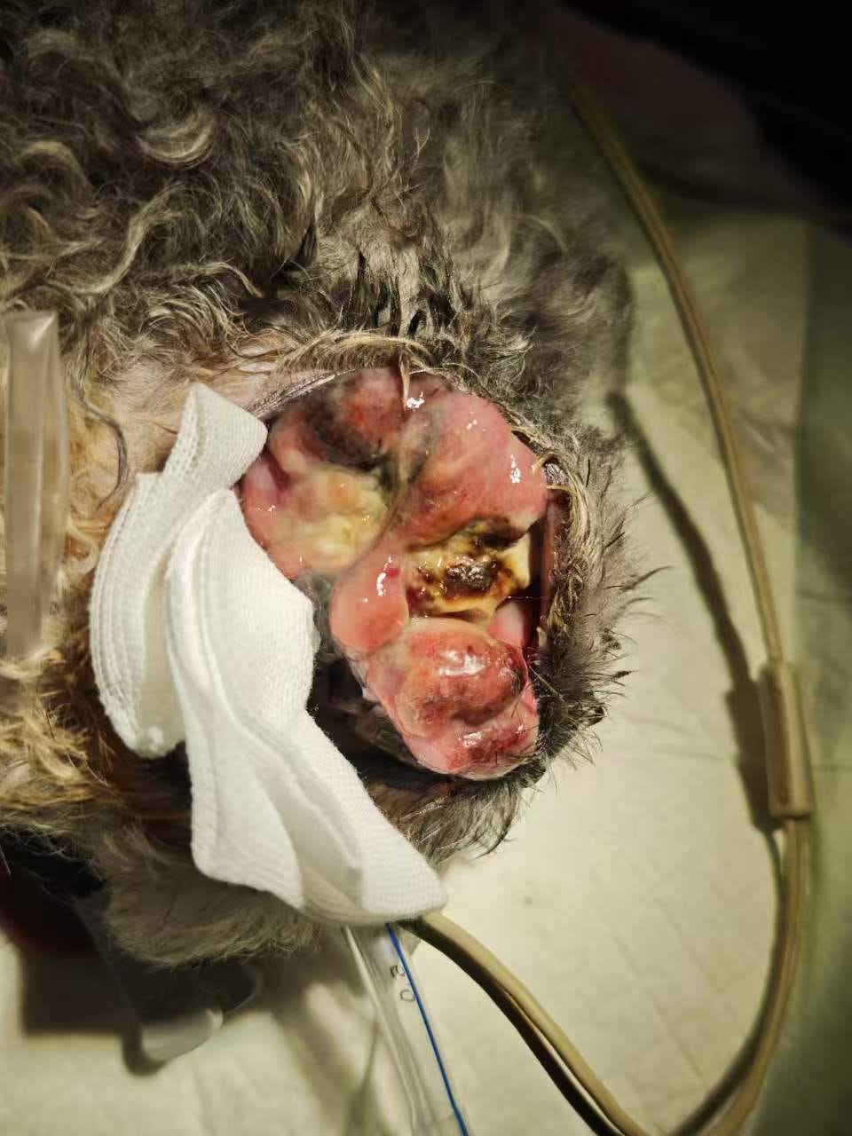
Figure 3. 27 August, after anaesthesia and immediately before the second injection. The tumour contains multiple irregular cavities and areas of necrotic material.
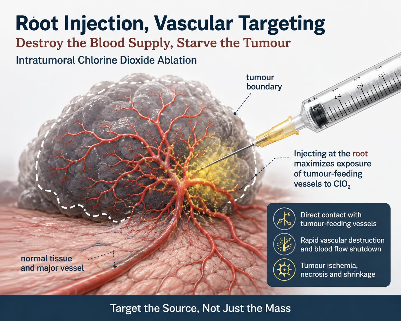
This time, injection placement was deliberately weighted toward the tumour root — the region where the tumour tissue connects with the surrounding body tissue. The procedural objective was not only to expose tumour tissue directly, but also to test whether delivery near accessible tumour-feeding regions could produce a broader vascular effect within the mass.
Approximately five minutes after injection, the gross appearance changed markedly. Large regions became dark brown to black and yellow-brown, with a clear change from the pre-injection appearance. As in the first procedure, the rapid colour and structural change is consistent with acute local tissue injury and necrotic change. Gross appearance alone, however, does not quantify the full depth or percentage of necrosis; follow-up imaging and tissue evolution remain important.
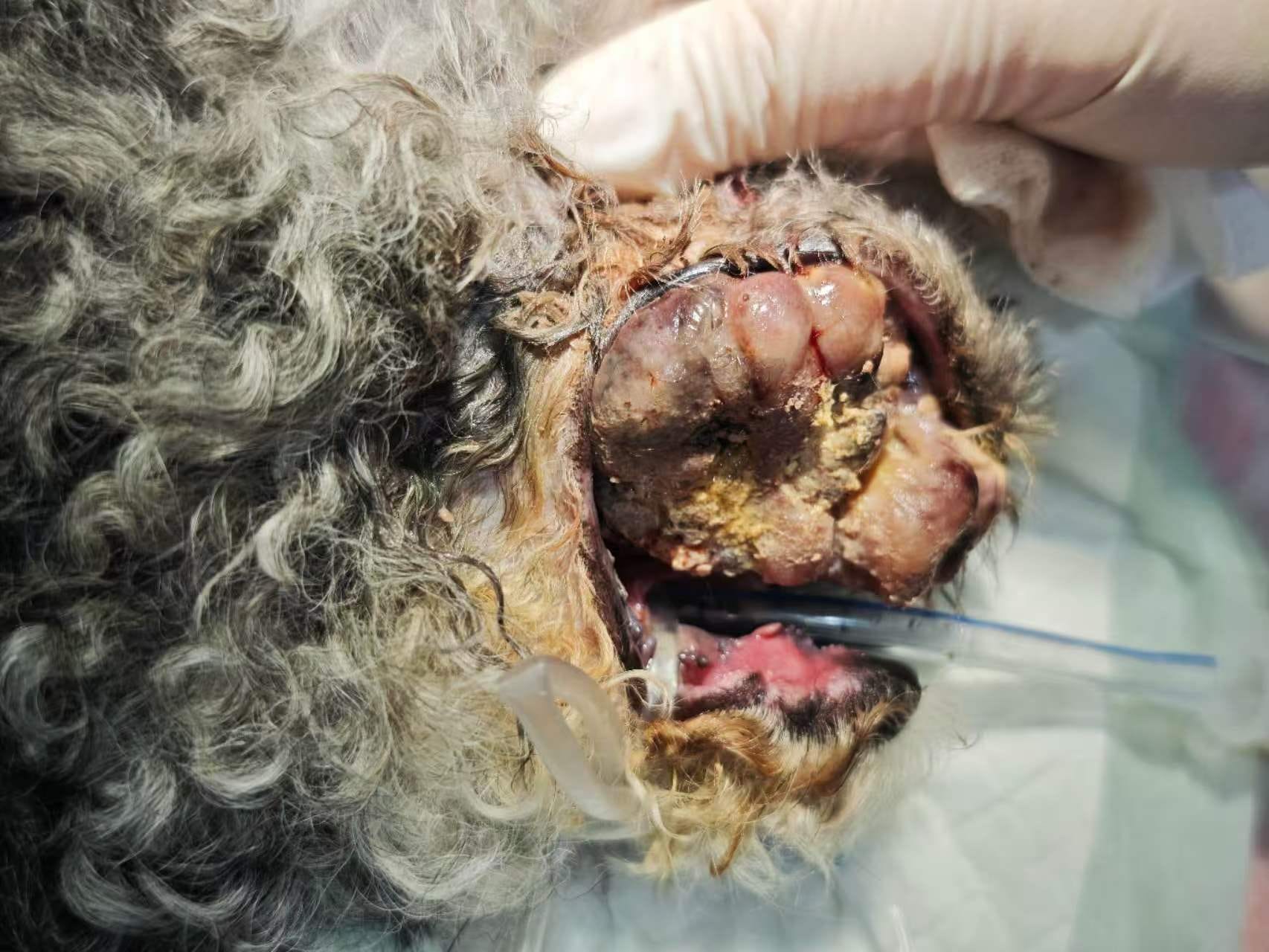
Figure 4. Approximately five minutes after the second injection. Marked darkening and yellow-brown change are visible across substantial portions of the treated mass.
