

Written for clinicians, veterinary radiologists, and researchers. This is a single-case imaging analysis and a treatment-planning discussion. It makes no efficacy claim, describes no treatment available to patients, and does not constitute medical or veterinary advice. All final decisions belong to the attending clinician.
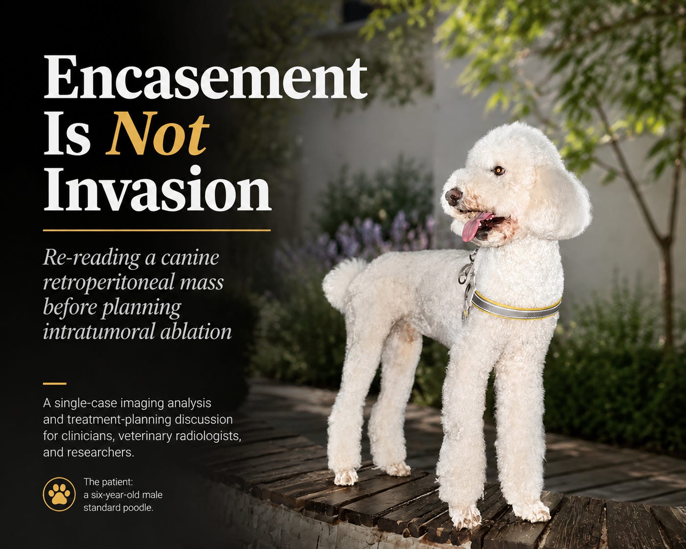
The patient: a six-year-old male standard poodle, photographed before the current episode.
The sentence that closes the door
A six-year-old male standard poodle presents with lameness. Contrast CT shows a large retroperitoneal mass in the sublumbar region. The report describes the liver, spleen, gallbladder, kidneys and bladder in careful detail, gives attenuation values for each of them, and then, for the one abnormal structure in the study, says that the mass invades the caudal vena cava and the aorta.
That sentence ends the case. No surgeon will open an abdomen to dissect a mass off a great vessel that a radiologist has already called invaded. The owner is told there is nothing to do. The dog is in pain, and the pain will get worse.
So it is worth asking what that sentence was based on. In this study, the answer is: loss of the fat plane between the mass and the vessels — and nothing else.
Fat-plane loss is the weakest sign in the entire vascular-involvement hierarchy. It has poor specificity. It occurs with simple apposition, with inflammation, with oedema, and with tumours that merely push. Every other sign that would actually establish invasion — luminal narrowing, luminal deformity, an intraluminal filling defect, a break in the wall contour, collateral circulation — was neither reported nor, as it turns out, present.
What the images allow you to measure
The study is a 0.625 mm acquisition, 854 slices, WL 40 / WW 350, with an on-screen scale bar. That combination is more informative than it looks: the scale bar gives real distances, the window settings let 8-bit display values be converted back to Hounsfield units, and the slice index gives craniocaudal extent directly as (slice difference x 0.625 mm).
Working across nine slices from Im 386 to Im 538, the mass has these dimensions:
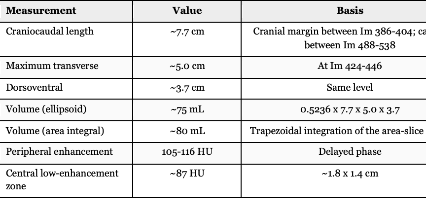
Two independent methods converge on roughly 75-80 mL. That number matters, because dose in intratumoral ablation is set as a fraction of tumour volume, and the report contained no volume at all.
The shape is not a sphere. Cross-sectional area rises steeply from the cranial tip, plateaus at 13-15 cm2 across Im 424-452, then falls away gradually toward the caudal end. This is a spindle: sharp cranially, blunt caudally, with the widest section in the cranial third and a longer caudal taper than cranial one.

Figure 1. Inferred shape in lateral and cross-sectional view, with the proposed approach direction.

Figure 2. Cross-sectional area along the long axis, measured slice by slice.
Three divergences from the original reading
1. The vessel call
The single most useful thing about a long tumour lying along a great vessel is that the vessel itself can be measured above the tumour, where it is normal, and again inside the tumour segment. If the mass is compressing or invading, the calibre changes. If it is only displacing, the calibre does not.
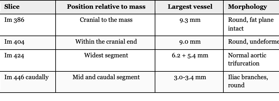
The aorta enters the tumour segment at 9.3 mm and is 9.0 mm inside it. The apparent narrowing further caudally is the normal trifurcation at L5-L6, not compression — a distinction that is easy to get backwards if you only scroll and do not measure.
Alongside that: no vessel cross-section on any slice is flattened or deformed; there is no intraluminal filling defect anywhere; there is no collateral circulation; both ureters are patent and opacified with no pyelectasia or hydroureter, despite one of them being circumferentially surrounded by tumour on a mid slice; and the colon is displaced but not narrowed.
A ureter has a far thinner and more compressible wall than an aorta. A tumour that fully surrounds a ureter without obstructing it is not a tumour that is compressing the aorta. The reading that fits the whole picture is displacement with segmental encasement, and no evidence of mural invasion.
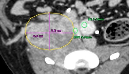
Figure 3. Im 404, cranial end of the mass: tumour outline with aortic and caval calibre.
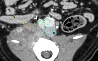
Figure 4. Im 386, beyond the cranial margin: vessels round, fat planes intact — the normal-calibre reference.
2. The mass itself was never characterised
The report gives attenuation values for the liver, the spleen, the gallbladder and the bladder, and none for the mass. It gives no dimensions and no volume. Yet those are precisely the numbers that separate lymphoma from sarcoma from metastatic carcinoma, and they are the numbers any local treatment plan is built on.
Measured here, the periphery enhances to 105-116 HU and the centre sits at about 87 HU. That gradient is real, but the centre is not frank liquefactive necrosis — necrosis on a delayed phase runs 20-40 HU. Eighty-seven HU is a hypoperfused zone that still contains tissue. It should not be written off, and it should not be skipped when planning injection points.
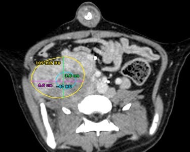
Figure 5. Im 452: outline, diameters, and regional attenuation.
3. The staging work-up stops short
The study is labelled thorax and abdomen. The report contains no thoracic findings at all — no lung windows, no nodule assessment, no mediastinal or thoracic nodal comment. For a malignant retroperitoneal mass, pulmonary metastasis screening is not optional; it determines whether local treatment is meaningful or purely palliative.
Three further omissions, all of them cheap to close: the prostate is not mentioned in a six-year-old intact-status-unknown male dog with a sublumbar mass and inguinal nodal involvement; neuter status and testicular position are not recorded, which matters because a retained abdominal testicular tumour can present exactly this way; and the lameness is never connected to the imaging, so nothing is said about the lumbosacral plexus, the external iliac vessels, or hindlimb oedema.
One more point, which cuts against my own reading as much as the original one. This is a delayed-phase study. In the delayed phase, intravascular iodine has largely equilibrated with soft tissue, and vessels are visible mainly because fat outlines them. Where the fat is gone, the vessel disappears — whether it is compressed, encased, or invaded. Every conclusion above about the vessels rests on calibre measurements and on indirect signs, not on direct visualisation of the vessel wall. An arterial and a venous phase, with reconstructions along the vessel axis, would settle the question outright.
Why the distinction changes the plan
Encasement is not a mild form of invasion. Anatomically it sits on the “not invaded” side of the line: the vessel wall and adventitia are intact, and there is a real tissue barrier between tumour and lumen. Operationally, though, encasement and abutment are completely different. Abutment can be worked around. Encasement cannot.
For this dog the consequence is specific. The best-perfused, most viable tissue is at the medial margin, facing the great vessels; the geometric centre is the hypoperfused part. So the tissue that most warrants treatment and the tissue that is safest to treat are not the same tissue. A staged plan does not dissolve that tension — it defers it. That has to be stated openly in the plan rather than glossed over as “gradual progress.”
What the method actually offers, and which parts transfer
Everything above is diagnostic. It establishes that the vessels are displaced rather than invaded, and therefore that a needle can be placed in this mass without the objection that killed the surgical option. It does not establish that anything worth doing follows. That depends on the properties of the agent, and on which of those properties survive the move from where they were observed to where they would be used here.
Intratumoral chlorine dioxide has six properties relevant to this decision. I list them, then separate them by whether they transfer to a deep, closed, retroperitoneal site.
The six properties
1. Dual mechanism. Chlorine dioxide oxidatively destroys tumour tissue directly, and separately destroys the microvasculature inside the tumour. It does not only kill what it touches; it also removes the blood supply sustaining the tissue downstream of what it touches.
2. Rapid necrosis. Marked tissue change appears in the treated zone soon after injection rather than over days.
3. Relatively little bleeding. Nothing is cut and no vessels are ligated, because the tumour is not removed from the body. Rapid destruction of tumour tissue and tumour vasculature has, in some cases, been accompanied by rapid disappearance of visible surface vascularity after treatment.
4. Relatively little inflammation. Necrosis occurs, but in the cases observed the local inflammatory response has not been commensurate with the speed and extent of that necrosis.
5. Broad-spectrum oxidative antimicrobial action. The agent is antimicrobial in its own right, independent of its effect on tumour tissue.
6. Repeatability. There is no requirement to resolve a large tumour in a single session. Treatment can be fractionated: reduce part of the mass, reassess, treat the remainder according to what the first session showed.
Which of these transfer to this case
Most of the observational base for these properties comes from superficial and ulcerated tumours, where the treated tissue is directly visible and where necrotic tissue has an existing route out of the body. This mass has neither. It sits in a closed retroperitoneal space with no communication to any surface. Applying the properties without adjusting for that would be careless, so:
— Property 1 (dual mechanism) transfers, and it is the property the plan depends on most. If killed volume can exceed contacted volume, then complete coverage is not a precondition of doing something useful, and a staged, partial-coverage approach is coherent rather than a compromise.
— Property 3 (little bleeding) transfers, and it is what makes the approach available at all here. The objection that closed the surgical option — a mass lying against the aorta and caudal vena cava — is an objection about dissection and haemostasis. A percutaneous needle placed from the lateral aspect does neither.
— Property 6 (repeatability) transfers, and without it there is no plan. A single-session treatment of an 80 mL mass adjacent to great vessels is not something anyone should attempt. Fractionation is only available if fractionation is actually possible.
— Property 4 (little inflammation) probably transfers, but this is inference, not observation. The relevant observations come from peripheral tumours, not from the retroperitoneum. It matters more here than anywhere, because inflammatory oedema in a closed space against great vessels would be a serious problem — which is exactly why it should be monitored rather than assumed.
— Property 2 (rapid necrosis) transfers as biology but not as a readout. In a superficial tumour the change is visible within minutes. Here nothing is visible, and imaging contour change in deep visceral tumours takes on the order of ten days. The necrosis may be just as fast; the operator will not know it for a week and a half.
— Property 5 (antimicrobial action) has little direct relevance. There is no open wound to protect.
