

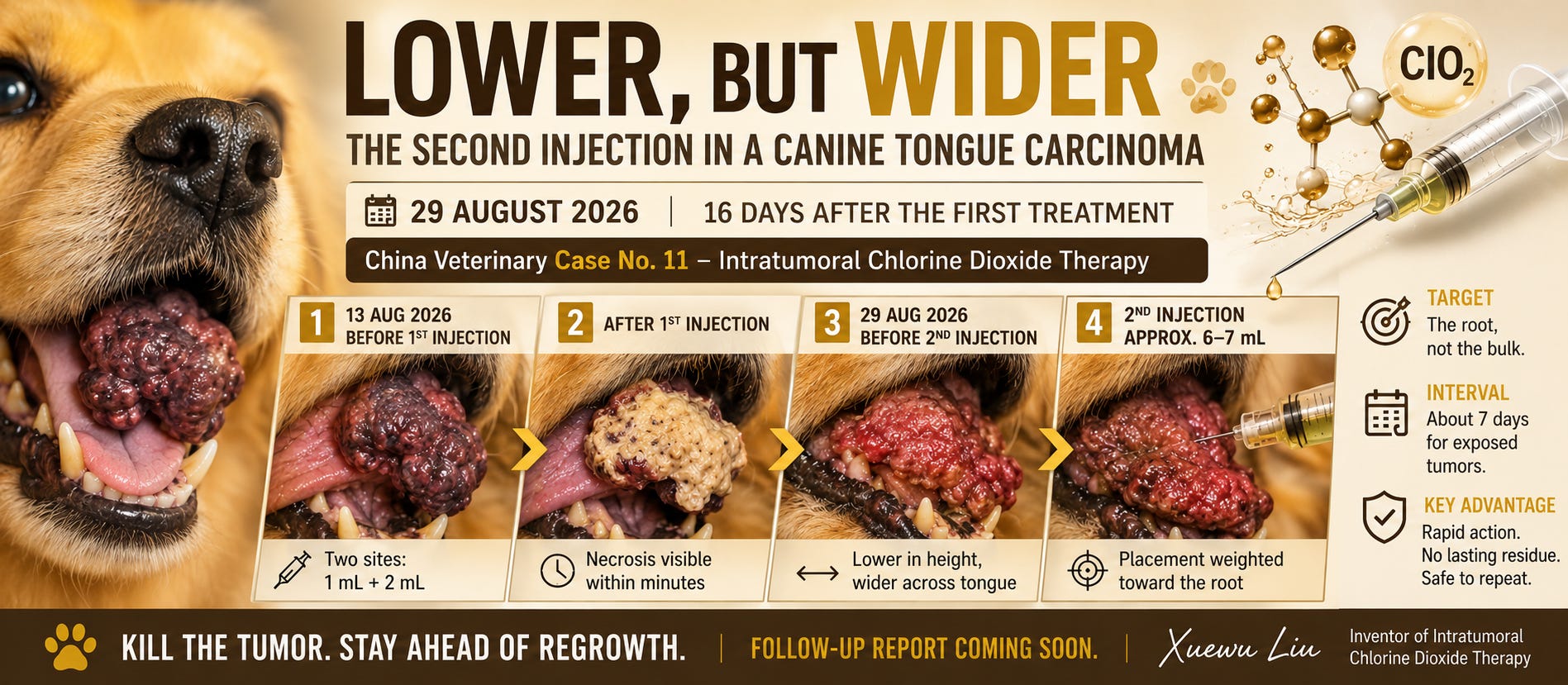
This is a follow-up to the 13 August 2026 report, When Chlorine Dioxide Changed a Tongue Tumor in Twenty Minutes, which documented the first treatment of a tongue squamous cell carcinoma in a 16-year-old mixed-breed dog of approximately 9 kg. That first procedure delivered only about 3 mL, split across two sites (1 mL and 2 mL), because the tumour was loose and larger boluses leaked straight back out of the surface. Twenty minutes later the treated tissue had turned pale yellow-white rather than black — the case that first showed us that blackening is a reading of blood, not a definition of the effect.
Today the dog came back for a second injection. What we found is, I think, more instructive than a clean success would have been.
Sixteen days later: the top died, the root did not
The tumour today is lower and wider than it was on 13 August.
That single sentence contains the whole lesson. The exophytic upper portion — the part that was directly reached by the first injection — underwent necrosis and was lost. The root, the region where the tumour meets the surrounding tongue tissue, remained viable and continued to grow, this time spreading laterally rather than upward.
The gross result is a mass that has lost height but gained width. The attending veterinarian could not confidently state whether total tumour volume had increased or decreased over the interval. That ambiguity is itself a finding: the volume killed by the first injection and the volume regrown in sixteen days were of roughly the same order. Necrosis and regrowth cancelled out, and the net change fell below the resolution of clinical estimation.
(Observation tier: the geometry change and the veterinarian’s inability to judge net volume are direct observations. The attribution of lateral growth specifically to a surviving root is a mechanistic inference — a well-supported one, but an inference.)
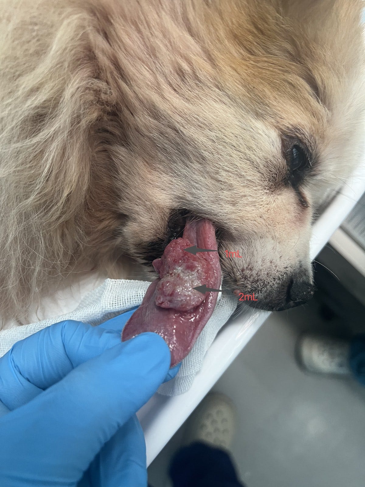
Figure 1. 13 August 2026, immediately before the first injection. The two arrows mark the planned injection sites and the volume delivered at each (1 mL and 2 mL). The tumour is exophytic, bulky in height, and loose in texture.
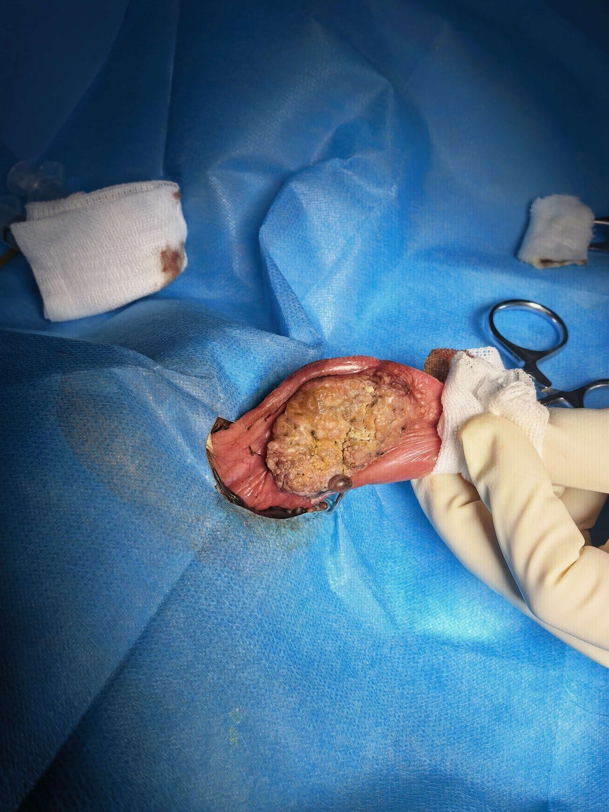
Figure 2. After the first injection. The treated tumour has become pale yellow-white and coagulated, sharply distinguishable from the surrounding viable tongue. Necrotic change was visible within minutes.
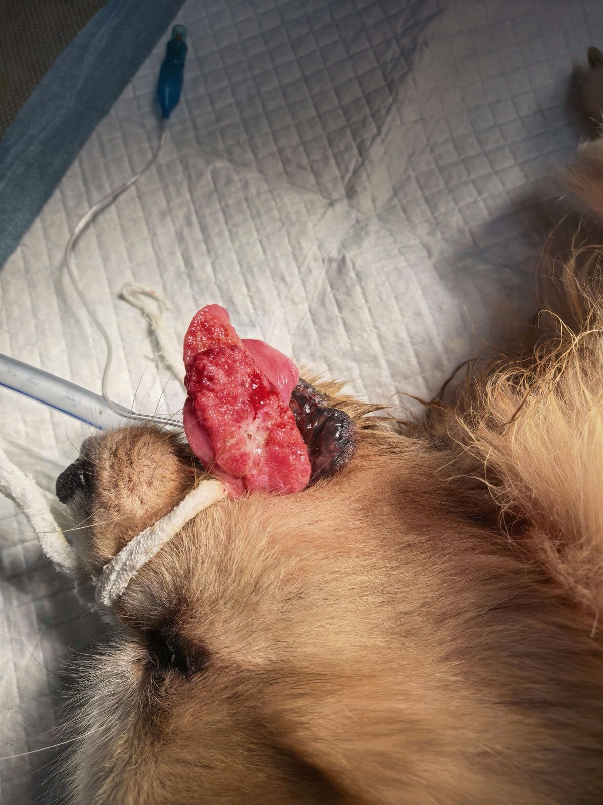
Figure 3. 29 August 2026, before the second injection. Compared with Figure 1, the mass is markedly lower in profile but broader across the tongue. The surface is red and granular rather than the pale, bulky mass seen sixteen days earlier.
The second procedure: easier, denser, and twice the dose
Three things differed from 13 August, and all three point the same way.
The injection was easier. On 13 August, the tissue was loose enough that every attempt at a larger bolus produced immediate surface leakage, forcing the veterinarian to fall back on multiple small-volume sites. Today the residual tumour felt comparatively dense, and it accepted fluid rather than expelling it.
The delivered volume roughly doubled. Approximately 6–7 mL was injected today, against about 3 mL at the first procedure — into a tumour that is, if anything, no larger than before. The retained fraction is presumably higher still, since leakage was much less of a problem.
Placement was weighted toward the root. Before the procedure I asked the veterinarian explicitly to prioritise coverage of the base — the junction between tumour and normal tissue — rather than the accessible bulk. This was a deliberate change of technique, not an incidental variation.
The veterinarian’s own feedback afterwards was that the injection volume was larger and that the visible signs of necrosis appeared faster than the first time, and on that basis he expects this round to produce more necrosis than the first.
(This is a prediction, not a result. It is testable within days, and I will report the outcome whether or not it holds.)
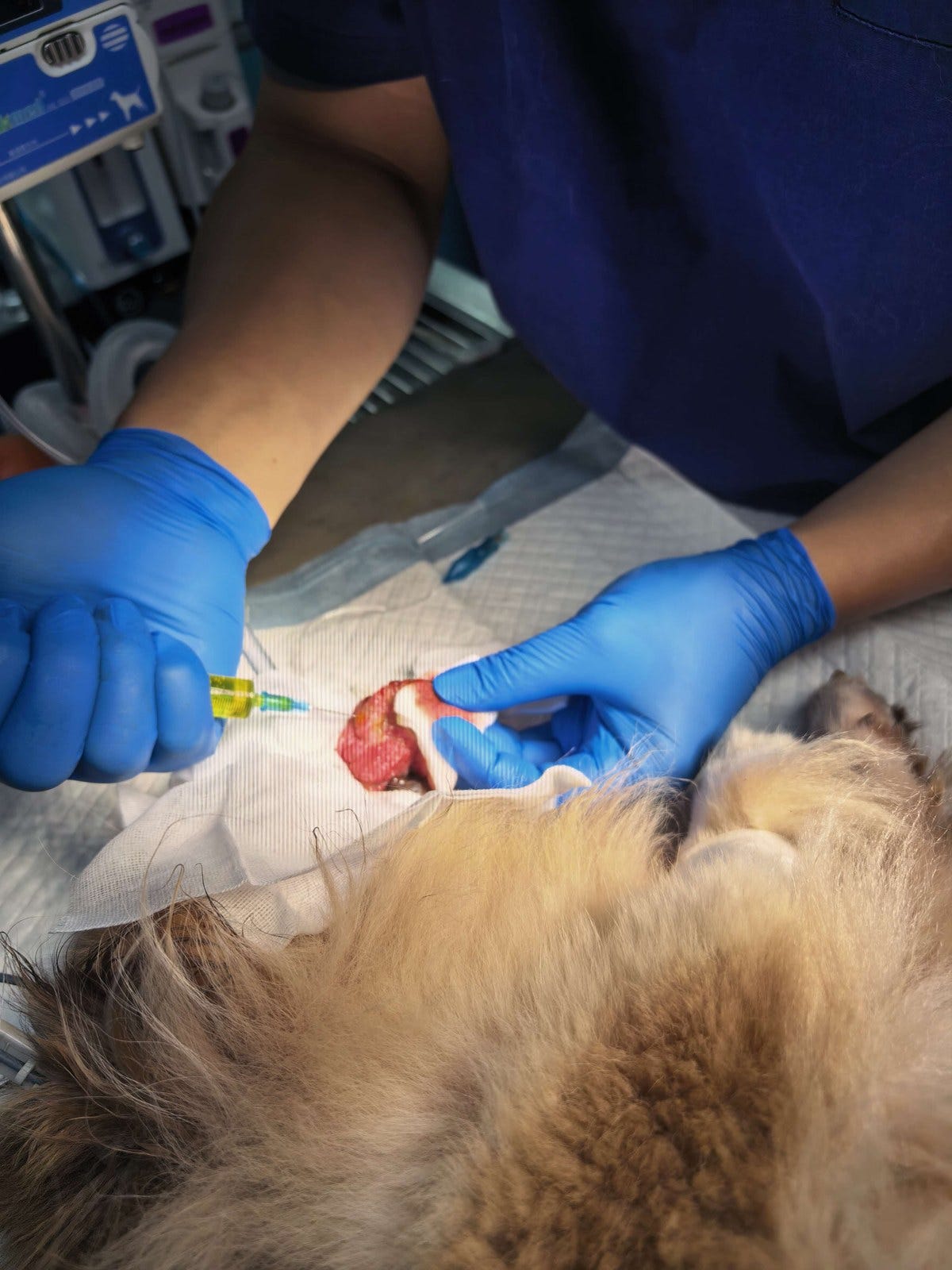
Figure 4. 29 August 2026, during the second injection. Approximately 6–7 mL of chlorine dioxide ablation solution was delivered, with placement weighted deliberately toward the tumour root.
Intratumoral chlorine dioxide injection, 29 August 2026
