Intratumoral therapy is having a moment, and the reason is not sentimental.
Systemic therapy has been the center of oncology for fifty years, and in solid tumors its progress has become expensive and incremental. Checkpoint inhibition transformed a minority of patients and left the majority where they were. Each new agent buys weeks against a background of toxicity that is distributed across the whole body in order to reach a lesion that occupies a few cubic centimeters of it. That arithmetic has not changed in a decade, and people have noticed.
Local therapy inverts the arithmetic. Put the agent where the tumor is, and the dose that matters at the target is achieved without the dose that matters everywhere else. The idea is old — the first attempts date to the nineteenth century — but it has returned because interventional imaging now makes almost any lesion reachable with a needle, and because the field has come to accept that a local intervention can have systemic consequences.
So the interest is real. What interests me more is the list of problems that comes with it.
The practitioner’s list
In October 2022, Eric Wehrenberg-Klee (Massachusetts General Hospital) and Rahul Sheth (MD Anderson) published a practical guide in Endovascular Today for interventional radiology teams preparing to take on intratumoral therapies — Intratumoral Therapies: How to Prepare Your Team for the Coming Wave. It is not a review of mechanisms. It is a description of what actually happens in the procedure room, written by two people who do it. That makes it more useful than most reviews.
Their list, condensed:
Scheduling. Many of these agents require weekly or biweekly injection, continuing for as long as the patient responds and residual tumor remains. Visceral injection increasingly requires sedation and CT guidance. This consumes procedural capacity that busy departments do not have.
Preparation windows. Several agents have only a short stability window after pharmacy preparation. A delayed procedure start can force a new preparation.
Operator continuity. Frequent injections make it difficult to keep one interventionalist with one patient. A team is required, which creates a communication problem: with large lesions treated by a clock-face approach across several sessions, every operator needs to know which sectors have already been covered. The authors also raise a quieter concern — that in this model the interventional radiologist risks being perceived as a functionary rather than a physician responsible for the patient.
Biohazard handling. For viral agents: contact precautions, pregnant staff excluded from care, the room cleaned with bleach afterward, and an occlusive dressing over the injected lesion for several days.
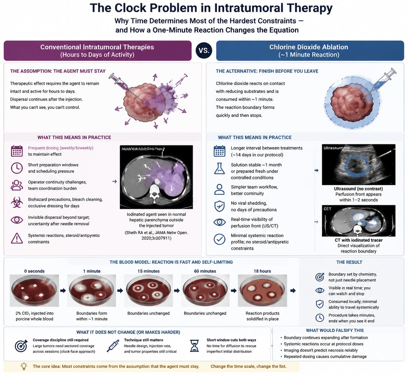
Delivery uncertainty. This is the part of the article I keep returning to. The authors observe that when we inject under imaging guidance, we assume the treatment goes where we put the needle, and that this assumption is often wrong. In the trials where the agent happened to be radiodense, MD Anderson physicians could see what normally cannot be seen: the therapy dispersed through a volume much larger than the tumor, despite correct needle placement. Their published figure shows an iodinated agent visible in normal hepatic parenchyma outside the injected lesion.
They also note that needle design and injection rate are under the operator’s direct control and materially affect distribution; that multi-side-hole needles distribute better than end-hole needles in preclinical work; and that these variables are not standardized across trials.
Adverse events. Reactions occur during injection or within about an hour, occasionally later: hypotension, hypoxia or subjective dyspnea, tachycardia, fever. Management is symptomatic, but steroids are to be avoided because they blunt the immune response the therapy is trying to provoke, and there is emerging concern that even acetaminophen may reduce response to checkpoint inhibitors.
That is an honest list, and I want to be clear that I am not pointing at it to say the field is doing something wrong. I am pointing at it because I think the items on it are not independent.
One assumption underneath
Read the list again and ask what each item presupposes.
The agent must remain in the tumor, intact and biologically active, for hours to days. Oncolytic viruses have to enter cells and replicate. Innate immune stimulators have to be present long enough to recruit and activate the cells that do the actual work. The therapeutic event is not the injection; the injection is the beginning of a process that unfolds on a biological timescale.
Once you accept that premise, most of the list follows from it:
If the agent persists, whatever escapes the tumor persists too — and escape is not hypothetical, it is the thing their iodinated figure shows. Persistence outside the target is what a systemic reaction profile is made of.
If the agent replicates, you need contact precautions, bleach, and days of occlusive dressing.
If the mechanism runs through the immune system, you cannot give steroids to treat the reaction the mechanism causes, and you have to worry about antipyretics.
If the mechanism requires immune priming, one exposure is rarely enough, so you dose weekly and inherit the scheduling and operator-continuity problems.
And the invisibility of the delivered volume matters far more than it otherwise would, because dispersal continues after the operator has left the room. You cannot correct what you cannot see, and by the time you could have seen it, the procedure is over.
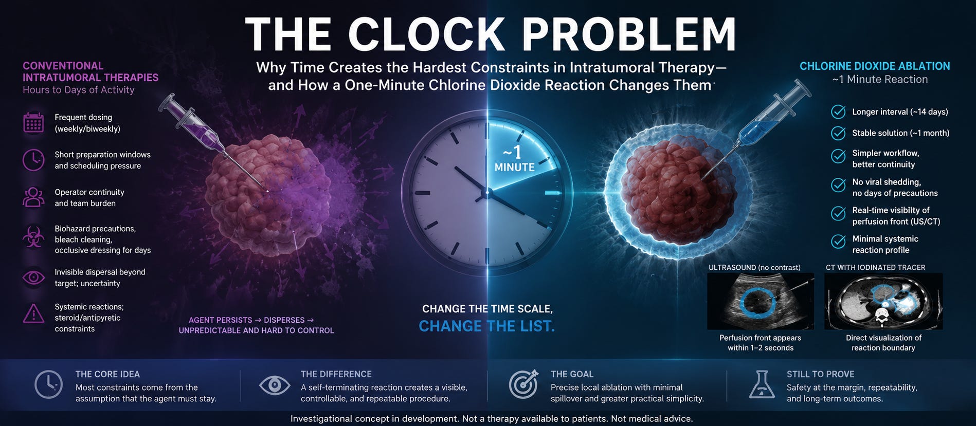
The unifying variable is time. Not chemistry, not immunology — duration. Almost every practical burden on that list is the cost of keeping something alive and active in a place for a long while.
Which raises a question the field has not really asked: what would change if the agent finished its work before the operator finished the procedure?
A seconds-scale agent
I have spent about fifteen years developing an intratumoral chlorine dioxide ablation system. In the United States it is being developed as a drug, under CDER — a 2% chlorine dioxide ablation agent injected into the tumor under image guidance, using a standard commercial syringe, with no proprietary delivery device. A pre-IND submission was filed on 15 August 2026; an orphan drug designation request for recurrent glioblastoma is under review.
The relevant property here is not potency. It is speed.
Chlorine dioxide is a selective oxidant. On contact with reducing substrates — and tissue and blood are nothing but reducing substrates — it reacts and is consumed. The reaction is not slow, and it does not wait.
We ran a simple experiment to characterize this. Into 500 g of chilled liquid porcine whole blood, we introduced discrete volumes of 2% (20,000 ppm) chlorine dioxide at six separate points, from 0.3 mL to 5 mL. Within one minute, each point had formed a sharply bounded reaction zone. At fifteen minutes the zones were essentially unchanged. At thirty minutes, unchanged. At sixty minutes the boundaries had not expanded — some radial striation appeared at the margins, which we have not explained — and eighteen hours later the reaction products had solidified in place.
The boundary forms in about a minute and then stops. The agent does not spread further because there is no agent left to spread. It is an ordinary result, and it is the whole argument. The footage and the full set of measurements are here: When Chlorine Dioxide Meets Whole Blood.
Two things happen inside that boundary, both fast: direct oxidative destruction of tumor cells, and destruction of the tumor microvasculature with loss of perfusion. The second matters as much as the first, because it means the treated volume does not need to be perfectly covered by the injected liquid to be devascularized.
And the same chemistry that produces the effect terminates it. Consumption is not a side condition of the safety argument; consumption is the safety argument.